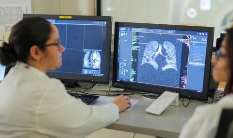
When you see your doctor for a health problem, you want a fast, accurate diagnosis. University of Missouri Health Care offers the latest diagnostic imaging services in central Missouri. We have medical technology and capabilities you can only find at an academic health center.
Our skilled radiology specialists have the knowledge and resources to give your doctor results quickly. That’s important to you because it means your doctor has detailed information as soon as possible to diagnose your health problem and begin treatment. Each provider has their routine for how results are relayed and our patient portal is another option our patients have for viewing their imaging results. Results are available online seven days after the exam is complete.
As a patient, you’ll have access to a full range of diagnostic imaging services including CT, MRI, 3T MRI, diagnostic, ultrasound, nuclear medicine and interventional radiology. We offer the most complete imaging services in the region.
Advanced imaging technology and expertise
The MU Health Care imaging team works closely with your primary care doctor to diagnose a health problem and develop a personalized treatment plan. Imaging technology is also used to guide a range of procedures such as biopsies (tissue samples), ESI injections, minimally invasive treatments to chemo embolizations.
Computed tomography (CT or CAT scan)
CT is a noninvasive procedure (doesn’t use surgery) that helps physicians diagnose and treat many medical conditions. CT images are acquired quickly using multi-row detectors. These images are viewed in a cross-sectional plane thus allowing the ability to see inside the organ or area imaged. It can be used to find infection, swelling, tumors or other problems throughout the body. Oral and IV contrast are sometimes administered to patients in order to assist the referring provider obtain a diagnosis. CT is used to image Code Stroke and Trauma patients upon arrival. For those patients who should not receive IV contrast, we also offer patients contrast enhanced ultrasound.
Magnetic resonance imaging (MRI) and functional magnetic resonance imaging (fMRI)
MRI uses very powerful magnets and radio waves to create clear pictures of your head, spine or other parts of your body. This noninvasive technology makes images of your body’s soft tissue, which includes organs, muscles and blood vessels. It can be used to identify healthy tissue and diseased tissue. Contrast enhanced ultrasound is also available for those patients unable to receive IV contrast.
FMRI utilizes an advanced MRI scanner to measure brain activity by detecting changes associated with blood flow. This technology is noninvasive and is used for diagnostic purposes.
MU Health Care offers standard MRI on 1.5T and 3T machines, as well as advanced imaging on the MAGNETOM Terra 7T MRI. There are fewer than 40 7T MRI scanners in North America, and MU Health Care is proud to be one of less than 10 health systems who have achieved accreditation through the American College of Radiology — the gold standard in medical imaging — for the 7T MRI.
Nuclear medicine
Nuclear medicine is a radiology imaging modality that uses radioactive isotopes to obtain images of the body’s physiology. In some cases, multiple day studies in which the patient leaves and returns at specific times are necessary. The radiation from the pharmaceuticals is generally minimal. Nuclear medicine exams are used to learn about the health of the heart, brain, lungs, gastrointestinal tract, skeleton and kidneys.
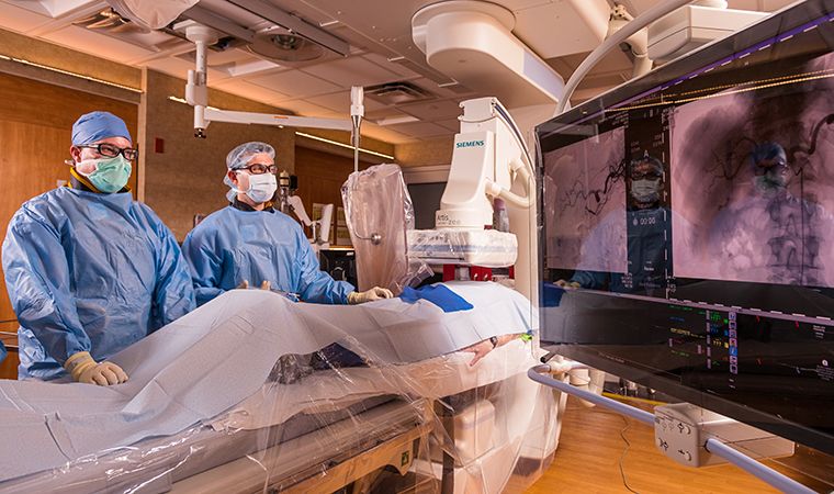
Interventional radiology (IR)
Interventional radiology is a subspecialty of radiology in which minimally invasive procedures are performed using image guidance (fluoroscopy/X-ray/CT/Ultrasound). IR is used for diagnostic and therapeutic care for a wide variety of vascular and non-vascular conditions. MU Health Care has specialists in heart care, neurology, cancer and other areas who use interventional radiology services for patients to unblock arteries, repair aneurysms and perform other advanced treatments such as embolization procedures (uterine artery, prostate, cancer tumors and more), angioplasty, stenting, shunting, imaging-guided drainage, and more.
Ultrasound
Ultrasound is a noninvasive imaging technique that uses high-frequency sound waves to create very detailed images of the body. Ultrasound is a painless test used to evaluate organs, tissues and blood vessels throughout the body using a transducer to send and receive high frequency sound waves that produce images. It can also be used to help guide biopsies. For patients unable to receive IV contrast, we do offer Contrast Enhanced Ultrasound Study (CEUS).
Fluoroscopy
Fluoroscopy emits radiation to provide real-time pictures of structures in the body that are moving, such as oral contrast as it goes from the esophagus to the stomach. It can be used to help find a foreign object in the body, place a needle for a medical procedure or adjust a broken bone.
X-ray
X-ray imaging, otherwise called plain film or general X-ray, are single shot pictures of specific anatomical regions as ordered by a physician. There are more general X-ray exams performed than any other exam. Our most common exam is a chest X-ray. We also do many upper extremity and lower extremity X-rays for broken bones.
Pediatric radiology
MU Health Care has a team of pediatric fellowship-trained radiologists to help provide a diagnosis while keeping your child’s mind — and yours — at ease. All imaging services are available for children, and some equipment is decorated specifically for kids to create a better experience for them.
Certain Children's Hospital imaging services are only performed on our main hospital campus at 1 Hospital Drive. These imaging services include CT scans, MRIs, nuclear medicine, fluoroscopy and interventional radiology.
Walk-in pediatric patients needing only a simple scan, such as a chest or foot X-ray, can still receive those services at any of our imaging locations.