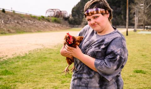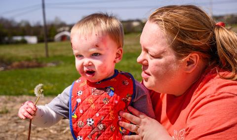
Achondroplasia is a form of disproportionate dwarfism that is characterized by shortened limbs (particularly in the upper arms and legs), enlarged head with frontal bossing, midface hypoplasia and bowed legs. There are fewer than 20,000 cases in the United States each year.
Achondroplasia is the result of a mutation in the FGFR3 gene and might be detected using radiological techniques, physical exams and genetic testing. Treatment of symptoms might include monitoring and surgery by doctors who specialize in skeletal dysplasia.
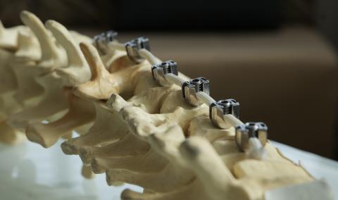
There is no cure for achondroplasia. Limb-lengthening surgery should only be discussed when the patient is old enough to weigh the risks and benefits. Adaptability products can help people with the condition do daily tasks such as hygiene, driving and reaching household items. Curvature of the spine can be treated with a brace or, less frequently, with spinal fusion surgery. Shoe lifts or inserts can address leg-length discrepancies. Surgery might be necessary to address leg alignment problems.
Achondroplasia heredity and genetics
The FGFR3 gene encodes the fibroblast growth factor receptor 3 on chromosome 4. Typically, FGFR3 is a membrane-spanning receptor that is active in slowing the production and development of cartilage. In achondroplasia, a single-point mutation in FGFR3 results in a virtually identical amino acid change of glycine to arginine, G380R. This modification causes fibroblast growth factor receptor 3 to be overexpressed, decreasing bone growth and causing disproportionate small stature. The mutant gene’s increased ability to slow bone growth is called a “gain-of function” mutation.
Achondroplasia is inherited in an autosomal dominant pattern, meaning only one mutant copy of the gene must be present to have the disorder. As a result, affected individuals have a 50% chance of passing the abnormal gene to their offspring. However, achondroplasia can also appear in individuals with no prior family history of the disease. In these situations, a spontaneous mutation was introduced. The risk of having another child with a spontaneous mutation resulting in achondroplasia is near zero. Achondroplasia gene abnormalities are fully penetrant, so any person with achondroplasia will show physical characteristics and symptoms of the disease.
Achondroplasia diagnosis and testing
A diagnosis can be obtained during fetal development after 22 weeks gestation. Routine ultrasound can detect the presence of short limbs, but an achondroplasia diagnosis is confirmed by testing the fetal DNA using amniocentesis. For families with a history of achondroplasia, amniocentesis or chorionic villus sampling (CVS) might be used to diagnose achondroplasia. However, not all individuals are diagnosed at birth, in which case clinical and radiologic evaluation by an experienced physician might be used to confirm achondroplasia.
Achondroplasia clinical features
- Short stature
- Mean adult height for men: 4-foot-3
- Mean adult height for women: 4-1
- Large head with frontal bossing
- Depressed nasal bridge
- Midface retrusion
- Short fingers, might have a trident-like appearance
- Joint contractures
- Genu varum
- Lordosis
- Kyphosis (particularly during infancy)
Achondroplasia radiographic features
- Decrease interpediculate distance of the lumbar spine
- Shortened tubular bones
- Flat, rounded iliac bones and horizontal acetabula
- Small sacrosciatic notches
- Radiolucent proximal femur and humerus
- Mild metaphyseal abnormalities
- Disproportionate humeral shortening
If an achondroplasia diagnosis is still in doubt after clinic and radiographic evaluations, genetic testing might be necessary.
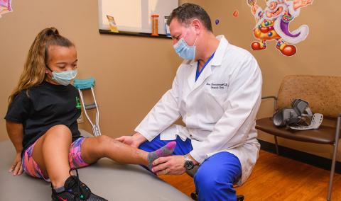
Achondroplasia-related orthopaedic problems and treatment
Growth monitoring
Growth should be monitored using achondroplastic growth, weight, weight-by-height and BMI charts. Limb-lengthening can be used to increase stature but should only be performed by surgeons trained in skeletal dysplasias. The patient should be old enough to make an informed decision regarding lengthening and weigh the benefits versus the risks involved.
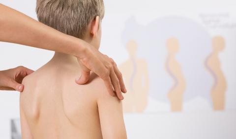
Kyphosis
Many infants develop outward curvature of the spine known as kyphosis because of weak muscle tone. As the infant grows and the core muscles become stronger, kyphosis usually disappears without requiring treatment. In these cases, the curve is flexible and tends to resolve when the child is laying face downward.
Persistent kyphotic curves occur in approximately 10%-15% of children with achondroplasia, in which case a clinical follow-up with spine X-rays is recommended. Treatment varies depending on the degree of kyphosis present, measured using a Cobb angle.
Kyphosis might be prevented by providing stable, supported seating of a child with achondroplasia. When the child is in a sitting position, angle the back at around 45 degrees to decrease the amount of weight pressing on the spine. Tummy time can also help spine stability.
Hypotonia
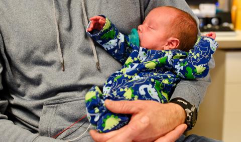
Mild to moderate hypotonia, or weak muscle tone, is common during infancy and will improve as the patient ages. Hypotonia and macrocephaly may make it hard for children to support their head. Delayed developmental milestones such as walking (18-24 months), crawling, standing, etc., may occur as a result of weakened muscle tone.
Genu varum
Bowing of the legs affects an estimated 60-80 percent of all children with achondroplasia and can arise from hypotonia, internal tibia torsion or twisting, and/or knee instability.
A physical exam should be used to monitor genu varum, searching specifically for a lateral thrust — the appearance that the knee suddenly jumps out to the side while walking. A series of X-rays called an extremity alignment series is also necessary to monitor the mechanical axis of the femoral head, knees and ankles.
Surgical intervention is typically not necessary but may be needed if the patient experiences extremity pain or if a lateral thrust is observed. Surgery should be performed by an orthopaedic doctor that specializes in skeletal dysplasia and may include derotational osteotomy or 8-plate.
Lordosis
Perhaps all children develop hyperlordosis and a prominent sacrum that tends to be parallel with the ground. In severe cases, it might result in chronic pain and require physical therapy to stretch the hip flexors and strengthen the core muscles. Bracing and surgical intervention are not necessary.
In adults with lordosis, surgical intervention may be necessary should the individual develop spinal stenosis with associated neurological symptoms.
Elbow contractures
Limitation in fully extending the elbows is common and usually ranges between 20 and 60 degrees shy of extension. As a result, adaptive devices might be necessary to aid in personal hygiene.
Joint hypermobility
Hypermobility, or loose joints, causes joint instability in the wrists and knees. When severe hypermobility is present, one might experience difficulty in fine motor activities such as knitting, writing, drawing, etc., and might find the hypermobile joints to fatigue more easily. Joint hypermobility is often noted at a young age then monitored as the child gets older. If symptoms interfere with everyday activities, consider modifying the environment with ergonomic keyboards, modified eating and writing utensils, etc.
Other common Achondroplasia problems and treatment
Weight management
Weight management is important for everyone, regardless of his or her stature. Going above a healthy weight increases the amount of pressure on the joints and can increase the risk of complications during surgeries or other medical procedures in addition to cardiovascular symptoms. When extra weight becomes distributed over a smaller surface area like that seen in any skeletal dysplasia, these observations may be more dramatic or more noticeable. However, there may be less incidence of obesity-related type 2 diabetes.
Because Body Mass Index (BMI) takes height and weight into account, dwarfs tend to have a naturally higher BMI but can still be considered healthy for their size. Use achondroplasia- specific BMI charts to monitor appropriate height and weight ratios.
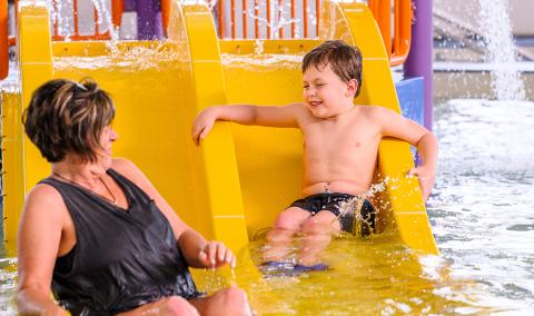
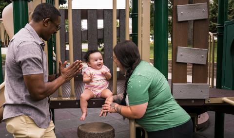
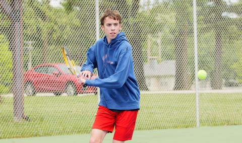
When exercising or engaging in physical activity, avoid high-impact sports such as football, wrestling, etc., unless the activity has been modified for achondroplasia (such as activities of the Dwarf Athletic Association of America). Low-impact and individualized sports and activities such as tennis, swimming, walking, etc., tend to put less pressure on the joints and are typically more comfortable forms of exercise.
Risk for hydrocephalus
Large head size is characteristic of those with achondroplasia. In children, this is accompanied with excess fluid in the brain, which may or may not need treatment. Symptomatic hydrocephalus presents in around 5 percent of children with achondroplasia.
Ventricle size and extra-axial fluid volume should be assessed in infancy using neuroimaging to establish a baseline. For the first year of life, head circumferences should be measured every one to two months and compared to an achondroplasia head circumference chart. After age 1, head circumference should be monitored with each regular check-up. Parents should be aware and check for signs of increased intracranial pressure. Symptoms include, but are not limited to, the following:
- Rapid increase of head circumference
- Seizures
- Irritability
- Vomiting
- Headaches
- Blurred or double vision
- Poor coordination
Risk for craniosynostosis
Although it is uncommon, children with achondroplasia may be more likely to experience premature closing of the skull sutures. This should be monitored by a pediatrician and referred to neurosurgery or a craniofacial surgeon if craniosynostosis is present.
Foramen magnum stenosis
The opening at the base of the skull is smaller and more constricted in all infants and children with achondroplasia, though risk of death from this complication is almost exclusively during the first year of life. As a result of foramen magnum stenosis, the respiratory system and upper cervical cord may become damaged. Respiratory system deficits can cause central sleep apnea. Infants should be assessed thoroughly to assess any neurological symptoms. If symptoms are present, neuroimaging should be completed and the foramen magnum size compared to achondroplasia-specific charts. If treatment is required, a subocciptal decompression is necessary to relieve the compression of the respiratory centers and nerves.
Parents should ensure their child has proper neck support when being held, as well as use a solid-back stroller and solid sleeping surface. Try to avoid umbrella strollers, baby swings/jumper swings, front or back carriers, and/or any other equipment that may cause neck instability.
Central sleep apnea
Central sleep apnea can be life-threatening and may occur because of foramen magnum stenosis or constriction. Unlike obstructive sleep apnea, the airway passages are open and functioning. However, the body is not receiving the signal from the breathing centers of the brain to inhale. People with central sleep apnea experience periodic episodes where breathing ceases altogether or breathing is shallow and not effective for appropriate respiratory activity. A polysomnography can be used to determine if the breathing problems result from airway obstruction or irregular respiratory signals from the brain. Daytime continuous or spot oximetry may also be necessary.
Treatment can include oxygen supplementation (used in a small percentage of patients) or a cervical decompression to treat foramen magnum compression.
Obstructive sleep apnea
Obstructive sleep apnea is common in children with achondroplasia between ages 2 and 10 and is a result of the inability to effectively breathe during sleep because of airway collapse. However, it is a lifelong issue in people with achondroplasia. It may be more likely because of small airways, redundant pharyngeal soft tissue and enlarged tonsils and adenoids. Obesity can also be a contributing factor. When left untreated, obstructive sleep apnea can cause chronic low levels of oxygen in the blood, high blood pressure and heart problems.
Snoring does not always signal significant airway obstruction. For this reason, monitoring additional symptoms of obstruction is appropriate. Symptoms may include:
- Self-arousals
- Loud snoring
- Nighttime vomiting
- Daytime irritability (or other signs of poor sleep)
- Bed-wetting
- Waking up gasping or choking
If serious obstructive sleep apnea is suspected, then polysomnography should be obtained for further information. There are several options for treatment, depending on the severity of the apnea:
- Adenoidectomy and/or tonsillectomy
- CPAP machine use
- BIPAP machine use
Ears and hearing
Middle ear dysfunction and/or conductive hearing loss is common in individuals with achondroplasia. If not treated from an early age, language and speech developmental delays may occur. Middle ear dysfunction is often unable to be managed. Hearing tests (behavioral audiometric and tympanometric) should be completed around 9-12 months of age and then repeated approximately annually.
Myringotomy and pressure equalizing tubes should be used aggressively. Ventilating tubes, should the child need them, should be used until ages 6-8. After this age, the Eustachian tubes should be functioning well and ventilation tubes are usually no longer necessary.
Call 573-882-BONE to make an appointment, or view MU Health Care specialists who treat achondroplasia.
Related Conditions & Treatments
- Adolescent Medicine
- Aerodigestive Program
- Down Syndrome
- Emergency Care for Kids
- Feeding Tubes for Children
- Juvenile Diabetes
- Hyperbaric Oxygen Therapy
- Inpatient Pediatric Psychiatric Care
- Neonatology
- Pediatric Anesthesiology
- Pediatric Cancer
- Pediatric Cardiology
- Pectus Carinatum
- Pediatric Chest Wall Disorders Program
- Pediatric Dermatology
- Pediatric Development and Behavior
- Pediatric ENT (Ear, Nose and Throat)
- Pediatric Epilepsy
- Pectus Excavatum
- Pediatric Eye Care
- Pediatric Gastroenterology
- Pediatric Infectious Diseases
- Pediatric Inpatient Rehabilitation
- Pediatric Nephrology
- Pediatric Neurology
- Pediatric Neurosurgery
- Pediatric Orthopaedics
- Pediatric Plastic Surgery
- Pediatric Primary Care
- Pediatric Psychiatry
- Pediatric Pulmonary Medicine
- Pediatric Sleep Medicine
- Pediatric Surgery Services
- Pediatric Surgical Services
- Pediatric Urology
- Pediatric Vascular Anomalies
- Pediatric Weight Management
- Sickle Cell Disease